
Have you noticed a small, firm lump inside your cheek, on your gums, or on your tongue? One possible cause is an oral fibroma, a usually benign growth of fibrous connective tissue that can develop following repeated irritation or trauma. Although oral fibromas are often painless, they may become uncomfortable when chewing, speaking, or brushing your teeth.
Modern dental technology offers different approaches to removing certain soft-tissue growths. One option is laser dentistry, which may allow a dentist to remove a suitable oral fibroma with precision and reduced bleeding.
So, can laser dentistry be used for oral fibroma removal? Yes, in selected cases. However, the correct treatment depends on the location and characteristics of the growth, the type of laser available, and the need for a biopsy to confirm the diagnosis.
If you are considering oral fibroma treatment, understanding the procedure, benefits, limitations, and recovery process can help you make an informed decision.
What Is an Oral Fibroma?
An oral fibroma is a non-cancerous growth made up of fibrous connective tissue inside the mouth. It may develop when the oral tissues experience repeated irritation over time.
Common locations include:
- The inner lining of the cheeks
- The gums
- The tongue
- The inner surface of the lips
- Areas repeatedly bitten or irritated by dental appliances
An oral fibroma may appear as a small, round, smooth lump that is similar in colour to the surrounding tissue. Some lesions may look pale or become irritated due to repeated friction.
Common contributing factors include cheek biting, rubbing from dentures, irritation from sharp teeth, and other forms of repeated local trauma.
Although oral fibromas are generally benign, not every lump inside the mouth is a fibroma. Other conditions can produce similar-looking growths, so a dental examination is essential.
Can Laser Dentistry Be Used for Oral Fibroma Removal?
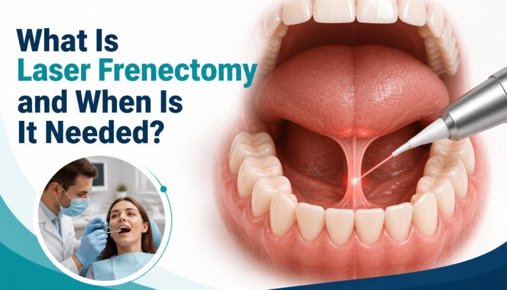
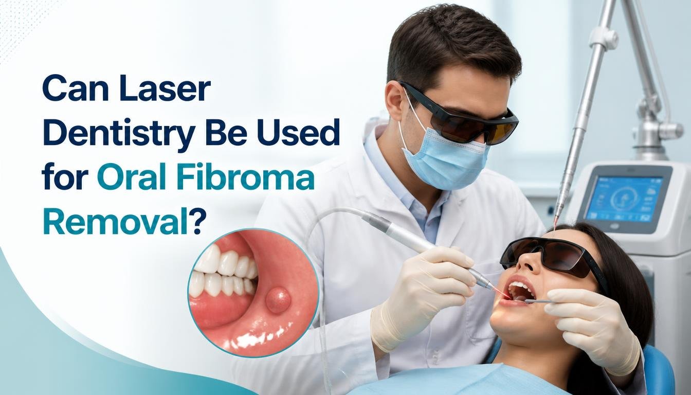
Yes. Laser dentistry can be used to remove certain oral fibromas through a procedure known as laser excision.
Dental lasers deliver focused energy to soft tissues, allowing trained dental professionals to cut or remove selected lesions. Published clinical reports describe the successful use of different laser systems for oral fibroma removal.
For example, a case report published in Compendium of Continuing Education in Dentistry in 2020 described the removal of an oral fibroma using an Er,Cr laser. A 2025 case series also reported promising outcomes using a 980 nm diode laser in four patients.
These findings support laser excision as a possible treatment option, but they do not establish that lasers are always better than conventional surgery. Larger studies are needed to compare outcomes across different lesions and treatment methods.
The dentist must first determine whether the growth is suitable for laser removal and whether the tissue needs to be submitted for laboratory examination.
How Does Laser Removal of an Oral Fibroma Work?
The procedure usually involves several steps.
1. Dental Examination and Diagnosis
The dentist examines the lump, checks its size and location, and asks about how long it has been present and whether it has changed.
Depending on its appearance and clinical history, the dentist may recommend a biopsy or removal followed by histopathological examination to confirm the diagnosis and rule out other conditions.
2. Treatment Planning
If removal is appropriate, the dentist selects a suitable technique. Laser treatment may be considered for certain soft-tissue lesions, while conventional surgical excision may be more appropriate in other cases.
The choice depends on the lesion, the equipment available, the clinician’s training, and whether an intact specimen is needed for diagnosis.
3. Local Anaesthesia When Needed
Local anaesthesia may be used to make the procedure more comfortable. The amount required varies according to the lesion and the treatment method.
Laser dentistry does not automatically mean that a procedure will be completely painless or that anaesthesia will never be necessary.
4. Laser Excision
The dentist carefully removes the lesion using the selected laser system and appropriate safety precautions.
Laser energy can help seal small blood vessels during treatment, which may reduce bleeding in suitable cases.
5. Tissue Examination and Follow-Up
When tissue is removed, it should be assessed according to the dentist’s diagnostic plan. Histopathological examination is particularly important when the diagnosis is uncertain or when the lesion has concerning features.
The dentist will also provide aftercare instructions and schedule follow-up if needed.
What Are the Benefits of Laser Dentistry for Oral Fibroma Removal?
Laser excision may offer several benefits in appropriately selected cases.
Reduced Bleeding
Some dental lasers help coagulate small blood vessels during soft-tissue procedures. This may improve visibility and reduce bleeding compared with certain conventional techniques.
Precision During Soft-Tissue Treatment
Laser energy can be directed at a specific treatment area. The actual precision and effect on surrounding tissue depend on the laser type, settings, and the clinician’s technique.
Potentially Less Postoperative Discomfort
Some patients may experience relatively mild discomfort following laser excision. However, pain levels vary, and laser treatment is not guaranteed to be more comfortable for every patient.
Potentially Easier Recovery
Selected cases have reported favourable healing after laser removal. Recovery depends on the lesion’s size, location, surgical technique, oral hygiene, and individual health factors.
Possible Reduction in the Need for Sutures
Some small soft-tissue lesions can be removed without stitches. Whether sutures are needed depends on the size and depth of the wound and the treatment performed.
Important: These potential benefits should not be interpreted as guarantees. Your dentist can explain which advantages are realistic for your particular case.
Laser Removal vs. Conventional Surgical Excision
Both laser excision and conventional surgical removal can be appropriate for oral fibromas.
| Feature | Laser excision | Conventional excision |
|---|---|---|
| Cutting method | Focused laser energy | Surgical blade or scalpel |
| Bleeding | May be reduced in suitable cases | Bleeding is managed using standard surgical methods |
| Anaesthesia | May be required | Commonly used |
| Stitches | May not be needed for some small lesions | May be needed, depending on the wound |
| Tissue diagnosis | Specimen suitability must be considered | Often allows collection of an intact specimen |
| Best option | Depends on the lesion and clinical assessment | Depends on the lesion and clinical assessment |
Neither approach is automatically the best choice for every patient. The priority is complete and appropriate removal, an accurate diagnosis, preservation of healthy tissue, and proper follow-up.
Is Laser Removal of an Oral Fibroma Safe?
Laser removal can be an appropriate option when performed by a qualified dental professional with suitable training and equipment.
However, all surgical procedures carry potential risks. These may include pain, swelling, bleeding, infection, delayed healing, scarring, or recurrence if the underlying source of irritation persists.
Another important consideration is diagnosis. A growth that resembles a fibroma may represent a different condition. Laser treatment must not replace the necessary diagnostic assessment or interfere with appropriate tissue examination.
Seek prompt dental evaluation if a mouth lump grows rapidly, bleeds without an obvious reason, develops an ulcer, becomes persistently painful, changes colour, or does not heal.
How Long Does Recovery Take After Oral Fibroma Removal?
Recovery varies according to the size and location of the lesion and the technique used.
After treatment, your dentist may recommend that you:
- Follow the specific oral-care instructions provided by your dentist.
- Choose soft foods if the treatment area feels tender.
- Avoid irritating the wound with hard, sharp, very hot, or spicy foods while it heals.
- Maintain gentle oral hygiene without disturbing the surgical site.
- Avoid smoking or tobacco products during recovery.
- Attend follow-up appointments and complete any recommended tissue examination.
Contact your dentist if you experience worsening pain, increasing swelling, persistent bleeding, fever, or other unexpected symptoms.
Can an Oral Fibroma Return After Laser Removal?
Yes, recurrence is possible, although it does not happen in every case.
Repeated trauma and irritation may contribute to the development of an oral fibroma. If the source of irritation remains, another lesion may develop in the same area or nearby.
Your dentist may recommend correcting contributing factors, such as a repeatedly irritating denture or a sharp tooth, where appropriate.
Follow-up is also important to check healing and review the tissue diagnosis. Any new or returning lump should be examined rather than assumed to be the same benign condition.
How Much Does Oral Fibroma Removal Cost?
The cost of oral fibroma removal in Noida depends on several factors, including:
- The size and location of the growth
- Whether laser or conventional excision is recommended
- The complexity of the procedure
- Whether a biopsy or laboratory examination is needed
- Follow-up and any additional treatment required
There is no single price that applies to every case. A dental consultation is the best way to obtain a treatment plan and an accurate estimate.
When comparing options, consider the dentist’s qualifications, diagnostic approach, and follow-up care rather than choosing a procedure based on price alone.
Where Can You Get a Dental Assessment for Oral Fibroma in Noida?
If you have noticed an unusual lump inside your mouth, arrange a dental examination to identify its likely cause and determine whether treatment is necessary.
The Dental Crowns offers dental services, including laser dentistry, at its clinic in Sector 79, Noida. Patients can discuss their oral health concerns with the clinic and ask whether laser treatment is appropriate for their particular condition.
Visit the Laser Dentistry in Noida page to learn more about the clinic’s laser dentistry services, or explore the complete dental services available.
Do not assume that every oral fibroma can be treated with a laser. A clinical examination is necessary to establish the diagnosis and select the most appropriate treatment.
Frequently Asked Questions
1. Can laser dentistry remove an oral fibroma?
Yes. Laser excision can be used for selected oral fibromas. The dentist must first assess the lesion and determine whether laser removal and appropriate tissue examination are suitable.
2. Is laser removal better than surgical removal for an oral fibroma?
Not in every case. Laser excision may reduce bleeding in selected cases, while conventional excision may be preferable when the lesion requires a particular surgical approach or intact tissue sampling. The best method depends on the diagnosis and clinical circumstances.
3. Is oral fibroma removal painful?
Local anaesthesia is often used when needed. Some patients experience mild postoperative discomfort, but the level of pain varies with the procedure and individual circumstances.
4. Does an oral fibroma need to be removed?
Not every lesion requires immediate removal. A dentist should examine the growth and determine whether monitoring, removal, or further investigation is appropriate. A suspected fibroma may need biopsy to confirm the diagnosis.
5. Can an oral fibroma become cancerous?
A typical oral fibroma is benign. However, other oral lesions can resemble a fibroma, so a proper examination and, when indicated, histopathological assessment are important.
6. How long does an oral fibroma take to heal after laser removal?
Healing time varies. A small published case series reported healing within seven to nine days after diode laser removal, but individual recovery may take longer.
7. Can an oral fibroma grow back after removal?
Recurrence is possible. Removing persistent sources of irritation and attending follow-up appointments may help manage contributing factors and identify any returning lesion.
8. Where can I consult a dentist for laser dentistry in Noida?
You can contact The Dental Crowns in Sector 79, Noida, to discuss your symptoms and ask whether the clinic offers an appropriate treatment option for your condition. Treatment suitability must be confirmed after examination.
Conclusion
Laser dentistry can be used for oral fibroma removal in selected cases and may offer benefits such as reduced bleeding and potentially less postoperative discomfort. However, it is not suitable for every lesion, and conventional surgical excision remains an important treatment option.
The most important first step is to have any persistent or unusual mouth lump examined by a qualified dental professional. An accurate diagnosis, appropriate removal technique, and necessary laboratory examination help ensure that the condition is managed correctly.
If you have noticed a lump inside your mouth, contact The Dental Crowns in Sector 79, Noida, to discuss your symptoms and arrange an assessment.
Book a dental consultation: The Dental Crowns
Medical disclaimer: This article is for educational purposes only and does not replace a professional dental examination, diagnosis, or individual treatment advice.