
When considering dental implants, one of the most important steps is understanding the condition and structure of your jawbone before treatment begins. A dentist may recommend a CBCT scan for dental implant planning to obtain detailed three-dimensional information that cannot always be seen clearly on conventional two-dimensional dental X-rays.
So, what role does a CBCT scan play in dental implant planning?
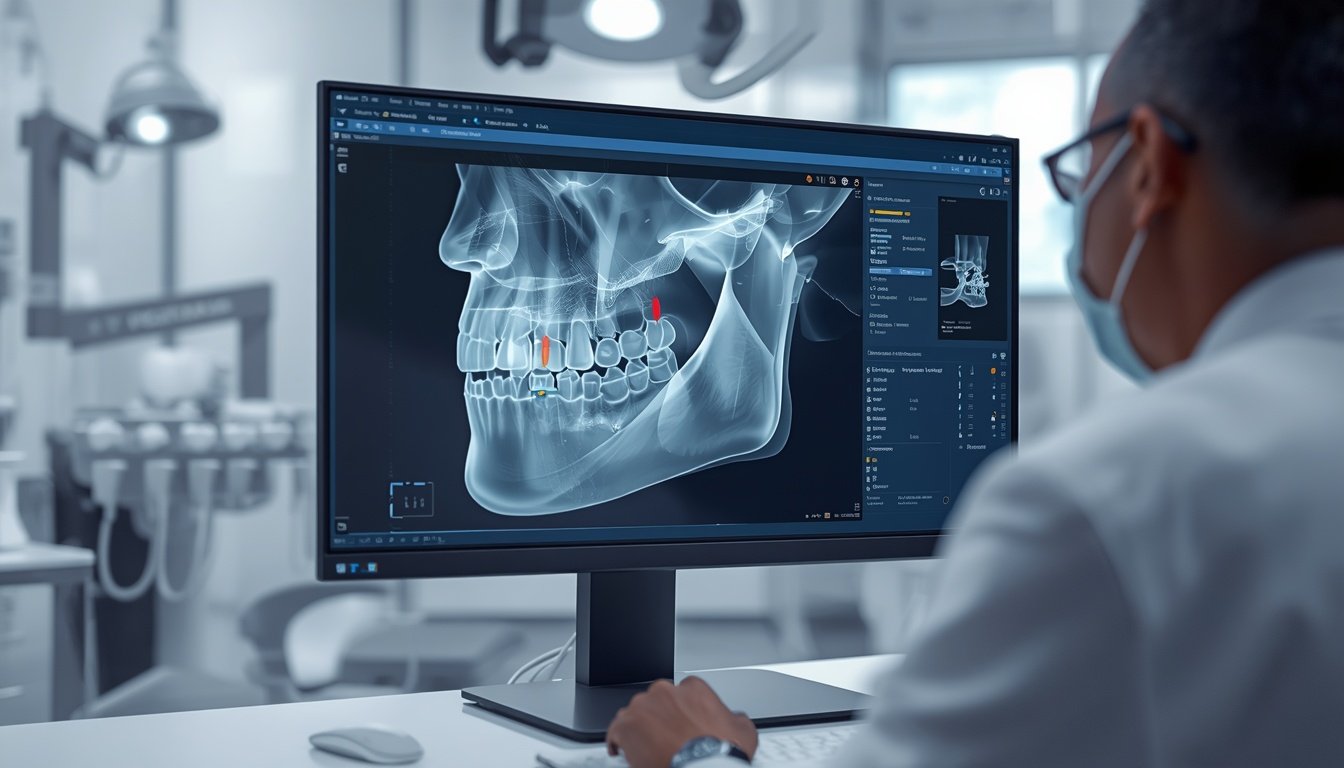
A CBCT, or cone-beam computed tomography scan, creates a 3D view of the teeth, jawbone and surrounding anatomical structures. This information can help the dental team evaluate the proposed implant site, assess available bone, identify important anatomical structures and develop a treatment plan based on the patient’s individual anatomy. The American Academy of Oral and Maxillofacial Radiology recommends cross-sectional imaging for implant-site assessment and identifies CBCT as the method of choice for obtaining this information.
For patients considering dental implants in Noida, understanding why CBCT imaging may be recommended can make the treatment-planning process easier to understand.
What Is a CBCT Scan in Dentistry?
CBCT stands for Cone Beam Computed Tomography. Unlike a conventional dental X-ray that produces a two-dimensional image, CBCT captures multiple X-ray projections and reconstructs them into a three-dimensional image of the area being examined.
According to the U.S. Food and Drug Administration (FDA), dental CBCT can provide 3D information about the teeth and oral and maxillofacial region and is used for several dental applications, including dental implant planning.
A CBCT scan can allow the dentist to examine:
- Jawbone width and height
- The shape of the alveolar ridge
- The position of nerves
- The location of the maxillary sinus
- The relationship between teeth and surrounding structures
- Potential bone deficiencies
- Anatomical variations
- The proposed implant site from multiple angles
This detailed information can be particularly useful when the implant site is anatomically complex.
Why Is CBCT Important for Dental Implant Planning?
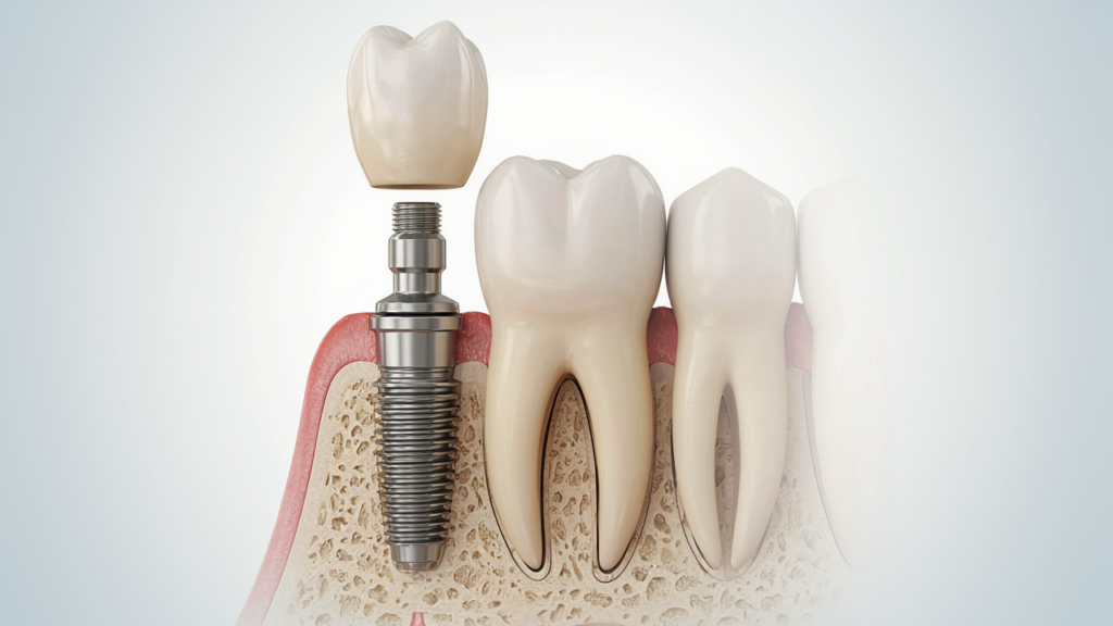
Dental implant placement is not simply about putting an implant where a tooth is missing. The implant must be planned according to the available bone, surrounding anatomy and the final restoration that will be placed on top of it.
Research published in the Journal of the American Dental Association describes how CBCT data can be used for virtual implant planning and can assist with transferring the planned implant position to the surgical site through a surgical guide.
Here are some of the key ways CBCT can contribute to implant planning.
1. It Provides a 3D View of the Implant Site
One of the biggest advantages of CBCT is three-dimensional visualization.
Traditional dental X-rays are useful, but a two-dimensional image can sometimes make it difficult to understand the exact width, depth and shape of the bone.
A CBCT scan allows the dentist to view the proposed implant site in multiple planes. This can provide a more detailed understanding of the patient’s anatomy before treatment.
The additional information may help the dentist determine whether the existing bone appears suitable for the planned implant or whether additional procedures may need to be considered.
2. It Helps Evaluate Available Bone
Adequate bone volume is an important consideration when planning a dental implant.
CBCT imaging can help assess the height and width of the available alveolar bone at the proposed implant site.
This information is important because the implant needs to be positioned within the available bone while also considering the final position of the replacement tooth.
A review published in PubMed notes that CBCT can provide detailed information about patient-specific anatomy, including the general condition of the jaw, bone quantity and other factors relevant to implant planning.
If there is insufficient bone, the dentist may discuss possible options such as bone augmentation or other site-development procedures where clinically appropriate.
3. It Helps Identify Important Anatomical Structures
The jaws contain important anatomical structures that need to be considered during implant planning.
For example, in the lower jaw, the inferior alveolar nerve canal may be relevant when planning implants in the posterior region.
In the upper jaw, the maxillary sinus may need to be evaluated, particularly when implants are being considered toward the back of the upper jaw.
CBCT can help the dental team visualize these structures in three dimensions and determine their relationship to the proposed implant site.
A clinical review of CBCT in oral implant dentistry describes its usefulness for evaluating the relationship between the alveolar crest and mandibular canal and for assessing structures such as the maxillary sinus and nasopalatine canal.
4. It Helps With Implant Position Planning
The ideal implant position is influenced by more than the missing tooth itself.
The dentist must consider factors such as:
- Available bone
- Adjacent teeth
- Anatomical structures
- Implant dimensions
- The position of the future crown
- The patient’s overall treatment plan
CBCT data can be combined with digital planning software to create a virtual representation of the proposed implant placement.
This approach allows the clinician to evaluate the planned position before surgery.
Research on presurgical implant-site assessment explains that CBCT combined with 3D software can improve visualization of bone, critical anatomy and restorative plans, supporting digitally guided implant planning.
5. It Can Support Computer-Guided Implant Surgery
CBCT is also an important component of some digital implant workflows.
After obtaining the 3D scan, the data may be imported into specialized planning software. Depending on the case and technology available, the clinician can virtually plan the implant position and potentially use that information to create a surgical guide.
A surgical guide can help transfer the digitally planned implant position to the clinical setting.
The Journal of the American Dental Association has described how CBCT data can support virtual planning and fabrication of surgical guides for implant placement.
However, the use of guided surgery is case-dependent and does not mean every implant patient requires the same digital workflow.
6. It May Help Identify the Need for Additional Procedures
Not every patient has the same amount or shape of available bone.
Some patients may have experienced bone loss following tooth loss, infection, trauma or other conditions.
In selected cases, CBCT can provide information that helps the dentist determine whether additional treatment may need to be considered before or alongside implant placement.
The AAOMR position statement specifically discusses CBCT in situations involving procedures such as sinus augmentation and bone grafting when clinically indicated.
This is one reason why CBCT can be particularly valuable in more complex implant cases.
7. CBCT Can Help With More Accurate Treatment Planning
A major goal of implant planning is to understand the patient’s anatomy before surgery rather than discovering important anatomical limitations during the procedure.
Three-dimensional imaging gives the dentist additional information that can be used alongside:
- Clinical examination
- Medical and dental history
- Conventional dental radiographs when indicated
- Digital impressions or scans
- The planned dental restoration
A PubMed-indexed review explains that CBCT can support presurgical diagnosis, treatment planning and the transfer of implant planning to surgical and prosthetic treatment.
This means CBCT should be viewed as part of a broader diagnostic process rather than as a replacement for a complete dental examination.
Is a CBCT Scan Required for Every Dental Implant?
Not necessarily.
The need for CBCT should be determined by the dentist based on the individual patient’s clinical situation.
The FDA states that dental CBCT should be performed when it is necessary to provide clinical information that cannot adequately be obtained through other imaging methods.
Similarly, professional recommendations emphasize that radiographic examinations should be justified based on the patient’s individual diagnostic needs.
For a straightforward implant case, conventional imaging and clinical examination may provide some of the required information. For more complex cases, cross-sectional imaging can provide additional anatomical information.
Therefore, a CBCT scan should not automatically be considered a routine test for every patient.
Is CBCT Safe for Dental Implant Planning?
CBCT uses X-rays, so radiation exposure is involved.
However, dental CBCT systems are designed for dental and maxillofacial imaging, and the radiation dose can vary depending on the machine, settings and field of view.
The FDA notes that dental CBCT generally delivers more radiation than conventional dental X-rays, while also emphasizing that CBCT should be used only when clinically necessary and with appropriate optimization.
The AAOMR also recommends using the smallest appropriate field of view and optimizing exposure parameters to obtain diagnostically acceptable images while limiting unnecessary radiation.
If your dentist recommends a CBCT scan, you can ask why the scan is needed and what information it will provide for your particular treatment plan.
What Happens During a Dental CBCT Scan?
A dental CBCT examination is generally quick and non-invasive.
The patient is positioned according to the imaging system being used. The CBCT unit rotates around the head while collecting multiple X-ray images. These images are then reconstructed into a 3D dataset.
The dentist or qualified imaging professional can then evaluate the images as part of the diagnostic and treatment-planning process.
The exact procedure can vary depending on the CBCT equipment and the area being scanned.
CBCT vs Traditional Dental X-Ray for Implant Planning
Both conventional dental X-rays and CBCT can have a role in dental care.
The main difference is the type of information they provide.
| Feature | Conventional Dental X-Ray | CBCT |
|---|---|---|
| Image type | 2D | 3D |
| Bone evaluation | Limited by projection | More detailed 3D assessment |
| Anatomical relationships | May be superimposed | Can be evaluated in multiple planes |
| Implant planning | Useful in selected situations | Particularly useful when cross-sectional information is needed |
| Radiation | Generally lower | Generally higher than conventional dental X-rays |
| Clinical use | Depends on diagnostic need | Should be clinically justified |
The AAOMR recommends cross-sectional imaging for assessment of dental implant sites and identifies CBCT as the preferred method for this purpose.
How CBCT Fits Into a Dental Implant Treatment Plan
A typical implant-planning process may involve several stages:
Step 1: Dental Consultation
The dentist discusses your missing tooth, oral health, medical history and treatment goals.
Step 2: Clinical Examination
The teeth, gums and surrounding tissues are examined to understand the condition of the mouth.
Step 3: Dental Imaging
Depending on the case, conventional X-rays and/or CBCT imaging may be recommended.
Step 4: 3D Assessment
If CBCT is required, the dentist can assess the available bone and relevant anatomical structures in three dimensions.
Step 5: Digital Treatment Planning
In suitable cases, CBCT information may be combined with digital planning tools to determine the proposed implant position.
Step 6: Implant Placement
Once the treatment plan is finalized, the implant procedure is performed according to the patient’s individual clinical requirements.
Step 7: Restoration
After the appropriate healing and integration period, the final restoration, such as an implant-supported crown, can be planned and placed according to the treatment protocol.
Why Personalized Implant Planning Matters
No two implant cases are exactly alike.
The location of the missing tooth, bone anatomy, adjacent teeth, gum condition, bite and overall oral health can all influence treatment planning.
This is why a CBCT scan should not be interpreted in isolation. It should be considered together with the clinical examination and other relevant diagnostic information.
For patients looking for dental implants in Noida, a personalized consultation can help determine whether 3D imaging is appropriate for their specific case.
You can learn more about Dental Implants at The Dental Crowns and explore the clinic’s other dental services.
CBCT and Dental Implant Planning in Noida
If you are considering replacing a missing tooth with a dental implant, the first step is a proper consultation.
The Dental Crowns provides dental implant treatment in Noida, with its clinic located at Civitech Stadia, Sector 79, Noida. The clinic’s implant service focuses on individualized treatment planning and implant solutions based on the patient’s needs.
Patients can also explore other dental treatments available at The Dental Crowns, including Painless Root Canal Treatment and other general, cosmetic and surgical dental services.
For questions about dental implants, CBCT imaging or your suitability for implant treatment, discuss your case directly with a qualified dental professional.
Frequently Asked Questions About CBCT and Dental Implants
1. What role does a CBCT scan play in dental implant planning?
A CBCT scan provides three-dimensional information about the jaw, bone and surrounding anatomical structures. This information can help the dentist assess the implant site and develop an individualized treatment plan.
2. Is CBCT better than a normal dental X-ray for implants?
CBCT provides 3D information, while conventional dental X-rays provide 2D images. CBCT can therefore provide additional anatomical information when cross-sectional imaging is clinically required. It does not necessarily replace conventional X-rays in every case.
3. Does everyone getting dental implants need a CBCT scan?
No. The need for CBCT depends on the individual case. The scan should be recommended when it is expected to provide clinically useful information that cannot adequately be obtained through other imaging methods.
4. Can CBCT show jawbone thickness?
Yes. CBCT can provide three-dimensional information that helps the dentist assess the dimensions and shape of the available bone at an implant site.
5. Can a CBCT scan show the dental nerve?
CBCT can help visualize important anatomical structures, including the mandibular canal containing the inferior alveolar nerve, which may be important when planning implants in the lower jaw.
6. Can CBCT help with sinus evaluation before an implant?
Yes. CBCT can provide information about the maxillary sinus and its relationship to proposed implant sites, which can be relevant when planning implants in the posterior upper jaw.
7. Is CBCT painful?
No. A CBCT scan is a non-invasive imaging examination and does not involve drilling, injections or surgical treatment.
8. How long does a dental CBCT scan take?
The actual scanning process is generally brief, although the total appointment time can vary depending on the equipment, area being examined and imaging protocol.
9. Does CBCT involve radiation?
Yes. CBCT uses X-rays. The FDA recommends that dental CBCT be used when clinically necessary and that exposure be optimized appropriately.
10. Can CBCT be used to plan guided dental implant surgery?
CBCT data can be incorporated into digital implant-planning workflows and, in suitable cases, may contribute to the creation of surgical guides.
11. Can CBCT determine whether I need a bone graft?
CBCT can provide information about the available bone and anatomy. Your dentist can use this information, together with the clinical examination, to determine whether additional bone augmentation or other procedures should be considered.
12. Should I ask my dentist why a CBCT scan is recommended?
Yes. Patients can ask what the scan is intended to evaluate and how the information will affect their treatment plan.
Final Thoughts
So, what role does a CBCT scan play in dental implant planning?
Its primary role is to provide detailed three-dimensional information that can help the dental team understand the implant site before treatment. CBCT can assist with evaluating available bone, identifying relevant anatomical structures, planning implant positioning and supporting digital or guided implant workflows when appropriate.
However, CBCT is not automatically necessary for every implant patient. The decision should be based on the individual patient’s clinical needs, with the examination appropriately justified and optimized.
If you are considering dental implants in Noida, speak with a qualified dentist for a complete evaluation and personalized treatment plan.
The Dental Crowns
Shop No. 16, 1st Floor, Civitech Stadia, Sector 79, Noida, Uttar Pradesh 201301
Phone: +91 89202 71787