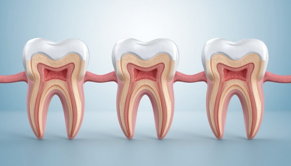


Have you ever wondered how dentists locate tiny or hidden root canals inside a tooth? During root canal treatment, identifying every canal is essential for removing infected tissue, cleaning the tooth properly, and preventing future complications. However, some root canals are extremely narrow, curved, calcified, or hidden behind complex tooth anatomy, making them difficult to detect with the naked eye.
Modern dentistry uses advanced diagnostic tools and specialized techniques to help dentists identify these hidden canals with greater precision. From digital dental X-rays and dental operating microscopes to 3D imaging technology, these methods help improve the accuracy of root canal procedures while preserving as much natural tooth structure as possible.
In this blog, we will explain how dentists find hidden root canals, why some canals are difficult to locate, and how advanced technology makes root canal treatment more effective.
What Are Hidden Root Canals?
A hidden root canal is a narrow or additional canal inside a tooth that may not be immediately visible during a routine dental examination or root canal procedure.
Every tooth has a different internal structure. While some teeth have a relatively straightforward root canal system, others may contain multiple canals, branches, curves, or additional anatomical variations.
For example, molars often have complex root canal anatomy. Sometimes, a tooth may contain an additional canal that is difficult to identify using conventional dental instruments.
If a canal remains undetected during treatment, infected tissue or bacteria may remain inside the tooth, potentially causing persistent pain, infection, or treatment failure.
Why Are Some Root Canals Difficult to Find?
Several factors can make root canals challenging to locate:
1. Complex Tooth Anatomy
Some teeth naturally have additional canals, branching pathways, or unusual root structures. These variations can make locating every canal more challenging.
2. Calcification of Root Canals
Over time, the pulp chamber and root canals may become narrower due to calcification. This process can occur because of aging, trauma, or other dental conditions.
3. Curved or Narrow Canals
Some root canals have sharp curves or very small openings. These canals may be difficult to identify and access using traditional instruments.
4. Previous Dental Treatment
Teeth that have undergone earlier dental procedures may have altered anatomy, restorations, or obstructions that make identifying hidden canals more complicated.
5. Limited Visibility
The internal structures of teeth are not directly visible during a routine dental examination. Dentists must rely on imaging, illumination, magnification, and specialized instruments to locate them.
How Do Dentists Find Hidden Root Canals During Treatment?
Dentists and endodontists use several diagnostic and clinical techniques to locate hidden root canals accurately.
1. Digital Dental X-Rays
Dental X-rays are among the first diagnostic tools used before starting root canal treatment.
Dentists may take periapical X-rays from different angles to examine the tooth’s roots, surrounding bone, and internal anatomy. Changing the angle can help reveal structures that overlap in a single image.
Digital dental X-rays help dentists:
- Identify the number and shape of roots.
- Estimate the length of root canals.
- Examine signs of infection around the root.
- Detect possible anatomical variations.
- Plan the root canal procedure.
Although dental X-rays provide valuable information, they produce two-dimensional images. Therefore, certain additional canals may remain difficult to identify.
2. Dental Operating Microscope
A dental operating microscope is one of the most valuable tools for identifying hidden root canals.
It provides powerful magnification and focused illumination, allowing dentists to examine the pulp chamber and locate tiny openings that might otherwise remain unnoticed.
Using a dental microscope, dentists can:
- Identify small or calcified canal openings.
- Detect additional canals in molars.
- Examine subtle anatomical details.
- Locate canal openings with improved visibility.
- Preserve healthy tooth structure during access preparation.
This technique is particularly helpful in complicated root canal treatments and cases involving calcified canals.
3. Cone Beam Computed Tomography (CBCT)
Cone Beam Computed Tomography, commonly known as CBCT, is an advanced dental imaging technique that provides three-dimensional views of teeth and surrounding structures.
CBCT may help identify:
- Additional or previously missed root canals.
- Unusual root canal configurations.
- Complex root shapes and curvature.
- Calcified canals and anatomical variations.
- Certain treatment complications.
Important: CBCT is not required for every root canal treatment. Dentists generally recommend it selectively when conventional examination and X-rays do not provide sufficient information for a complex case.
4. Ultrasonic Instruments
Dental ultrasonic instruments use high-frequency vibrations to help dentists carefully remove calcifications, debris, or obstructions around the pulp chamber.
These instruments can improve access to areas where hidden canal openings may be located.
When used under magnification, ultrasonic tips allow dentists to work with greater precision while minimizing unnecessary removal of healthy tooth structure.
This technique can be especially useful when locating calcified or obstructed canals.
5. Careful Examination of the Pulp Chamber
The pulp chamber is the central area inside the tooth where the root canals begin.
Dentists examine its floor, shape, color, and anatomical landmarks to identify the positions of canal openings.
Certain natural patterns and differences in dentin color can provide clues about the location of additional canals.
A careful and conservative access procedure is important because excessive removal of tooth structure can weaken the tooth.
6. Electronic Apex Locators
Electronic apex locators are specialized instruments that help dentists determine the working length of a root canal by detecting the position of the root canal file inside the tooth.
They are mainly used to determine canal length rather than directly discover hidden canals. However, they can support accurate treatment once a canal has been located and accessed.
Combining apex locators with radiographic examination can help dentists achieve more precise working-length measurements.
Can Artificial Intelligence Help Detect Hidden Root Canals?
Artificial intelligence is increasingly being explored in dental imaging and diagnosis.
AI-assisted dental software may help analyze radiographs and CBCT images, identify anatomical patterns, and highlight areas that require closer examination.
These technologies may provide additional support to dentists when evaluating complex root canal anatomy. However, AI is not a replacement for professional clinical judgment, diagnostic imaging, or the experience of an endodontist.
What Happens If a Dentist Misses a Root Canal?
If a root canal remains untreated during the procedure, bacteria and infected tissue may persist inside the tooth.
Possible consequences include:
- Persistent or recurring tooth pain.
- Continued infection inside the tooth.
- Swelling around the affected area.
- Development or persistence of an abscess.
- Delayed healing after root canal treatment.
- The need for root canal retreatment.
Not every unsuccessful root canal is caused by a missed canal. Other factors, including cracks, restoration problems, and persistent infection, may also affect treatment outcomes.
This is why proper diagnosis, careful treatment planning, and follow-up are important.
Does Finding Hidden Root Canals Make Root Canal Treatment Painful?
No. Locating hidden canals does not automatically make root canal treatment more painful.
Root canal procedures are generally performed under local anesthesia to keep patients comfortable during treatment. Advanced magnification and diagnostic technology can help dentists perform complex procedures more precisely.
Some tenderness or sensitivity may occur after treatment, but persistent or severe pain should be evaluated by a dentist.
When Should You Consult an Endodontist?
An endodontist is a dental specialist who focuses on diagnosing and treating problems involving the dental pulp and root canal system.
You may be referred to an endodontist when:
- A tooth has unusually complex root canal anatomy.
- Root canals appear calcified or difficult to locate.
- Previous root canal treatment has not healed as expected.
- A persistent infection is suspected.
- Additional diagnostic imaging or specialized equipment may be required.
Seeking an appropriate specialist can help manage complicated cases and determine the most suitable treatment approach.
Conclusion
Finding hidden root canals requires a combination of clinical expertise, detailed diagnosis, and modern dental technology. Digital dental X-rays, dental operating microscopes, CBCT imaging, ultrasonic instruments, and careful examination of the pulp chamber all play important roles in identifying complex root canal anatomy.
When a hidden canal is suspected, the dentist may use additional diagnostic methods or refer the patient to an endodontist for specialized care.
If you are experiencing persistent tooth pain, sensitivity, or signs of dental infection, consult a qualified dentist for an examination. Early diagnosis and appropriate treatment can help preserve your natural tooth and prevent complications.
For more information about dental procedures and treatment options, visit The Dental Crowns.
Frequently Asked Questions (FAQs)
1. How do dentists find hidden root canals?
Dentists use dental X-rays, magnification, microscopes, specialized instruments, and sometimes CBCT imaging to locate hidden canals.
2. Can a root canal be hidden on an X-ray?
Yes. Some canals may overlap with other anatomical structures or be too narrow to appear clearly on conventional two-dimensional X-rays.
3. What is the best technology for finding hidden root canals?
A dental operating microscope and selectively used CBCT imaging can be particularly helpful in complex cases.
4. Can CBCT detect missed root canals?
CBCT provides three-dimensional images that can help identify additional canals that may not be visible on conventional X-rays.
5. Why are root canals sometimes calcified?
Calcification may occur due to aging, dental trauma, or other changes inside the tooth, making canals narrower and more difficult to locate.
6. Can hidden root canals cause tooth pain?
Yes. If a canal contains untreated infected tissue, it may contribute to persistent pain, inflammation, or infection.
7. Can dentists treat calcified root canals?
Many calcified canals can be treated, but the difficulty depends on their location, degree of calcification, and tooth anatomy.
8. Does every root canal require a dental microscope?
No. Microscopes are particularly beneficial for complex procedures, calcified canals, and cases where additional precision is required.
9. What happens if a hidden canal is missed?
A missed canal may allow infection to persist and could lead to symptoms or the need for retreatment.
10. Is an endodontist better for complicated root canal treatment?
An endodontist has specialized training in diagnosing and managing complex root canal problems and may be appropriate for difficult cases.
11. Are additional root canals common in molars?
Molars often have more complex root anatomy and may contain additional canals, which is why dentists examine them carefully.
12. Is CBCT safe for root canal diagnosis?
CBCT uses X-rays, so it should be recommended only when clinically justified. Dentists consider its potential benefits and radiation exposure before prescribing it.