

Have you ever wondered how dentists treat the tiny, complex spaces inside your teeth with greater accuracy? Modern dentistry has introduced advanced technologies that help dental professionals examine areas that are difficult to see with the naked eye. One such innovation is the root canal microscope, also known as a dental operating microscope.
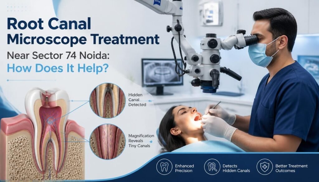
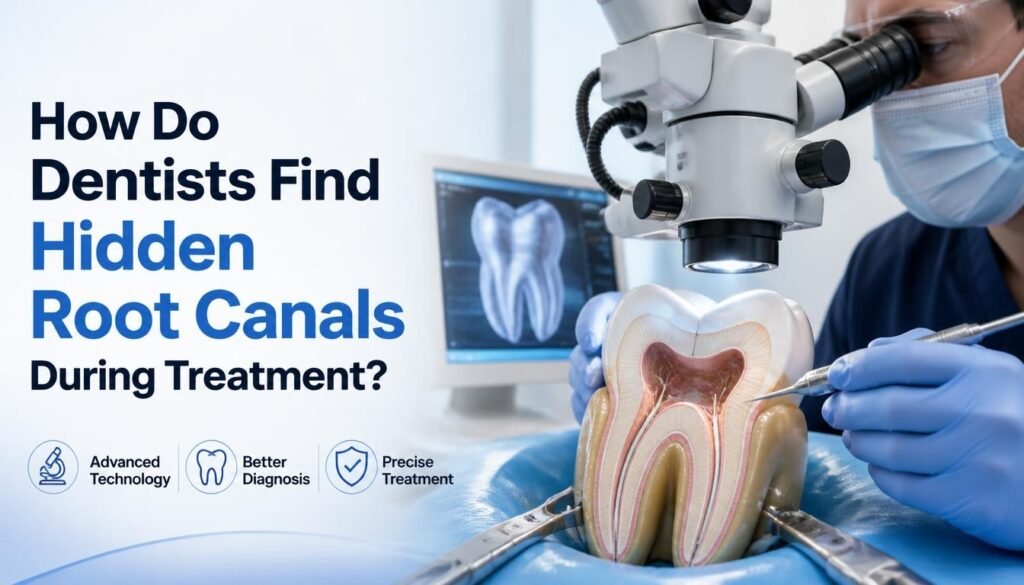
A root canal microscope is an advanced dental instrument that provides magnification and focused illumination, allowing dentists to examine the internal structures of teeth in greater detail. It can be particularly useful during complex root canal procedures, where identifying narrow, hidden, or curved canals is essential.
For patients considering root canal treatment in Noida, understanding how microscope-assisted dentistry works can help them make informed decisions about their dental care.
At The Dental Crowns, we believe patient education is an important part of comfortable and informed dental treatment. Let’s explore what a root canal microscope is, how it works, and why it is valuable in modern endodontics.
What Is a Root Canal Microscope?
A root canal microscope is a specialized dental operating microscope used during endodontic procedures, including root canal treatment and root canal retreatment.
Unlike traditional dental examination methods, which rely primarily on the dentist’s eyesight and conventional lighting, a dental operating microscope provides magnified visualization of the treatment area.
Depending on the equipment and settings, dental microscopes can offer magnification of approximately 4x to 25x, helping dentists observe minute structures inside the tooth.
The microscope typically consists of:
- Magnification lenses: Enlarge the treatment area for detailed examination.
- Focused illumination: Provide bright, concentrated light inside the tooth.
- Adjustable viewing system: Allows the dentist to change magnification according to the procedure.
- Ergonomic positioning: Supports a comfortable and controlled working posture.
This combination of magnification and illumination helps dentists perform delicate procedures with improved visibility.
How Does a Root Canal Microscope Work?
The root canal microscope works by magnifying the treatment area while directing focused light into the tooth.
During root canal treatment, the dentist needs to access the pulp chamber and locate the root canals. These canals may be extremely narrow, curved, or difficult to identify, particularly in molars.
A microscope helps the dentist observe the internal anatomy more clearly throughout different stages of treatment.
Step 1: Detailed Examination of the Tooth
Before treatment, the dentist examines the affected tooth and evaluates its condition. Magnification may help identify tiny cracks, structural defects, and other details that require closer inspection.
Step 2: Accessing the Pulp Chamber
Once the tooth is prepared for treatment, the dentist creates an access opening to reach the pulp chamber. The microscope helps visualize the chamber while supporting a more controlled approach.
Step 3: Locating Root Canals
Some teeth have additional or unusually positioned canals. Magnification and illumination can help the dentist locate these canal openings, including canals that might otherwise be difficult to detect.
Step 4: Cleaning and Shaping the Canals
The dentist cleans and shapes the root canal system using specialized instruments. Microscopic visualization can help guide delicate procedures and support the preservation of healthy tooth structure.
Step 5: Filling and Sealing the Tooth
After cleaning and disinfection, the root canals are filled and sealed. The tooth is then restored with an appropriate filling or dental crown, depending on its condition.
How Does a Root Canal Microscope Improve Treatment?
The primary advantage of a dental operating microscope is improved visibility. However, better visualization can support several important aspects of endodontic treatment.
1. Helps Locate Hidden Root Canals
One of the major challenges in root canal treatment is identifying every canal inside the tooth.
Some teeth, especially upper molars, may contain additional canals such as the second mesiobuccal canal (MB2). If a canal remains undetected, infected tissue or bacteria may persist inside the tooth.
A dental microscope helps the dentist examine the pulp chamber floor and identify canal openings with greater precision.
2. Improves Treatment Precision
Root canal treatment involves working within very small anatomical spaces.
Magnification allows the dentist to observe fine details while performing delicate procedures. This improved visualization can support more controlled instrument movements and accurate treatment.
3. Supports Conservative Tooth Preparation
Preserving healthy natural tooth structure is an important consideration during dental treatment.
With improved visibility, a dentist may be able to identify the required treatment area more precisely and avoid unnecessary removal of surrounding tooth structure.
This is particularly relevant when treating calcified or narrowed canals.
4. Helps Identify Calcified Root Canals
Over time, some root canals become narrower due to calcification. This can make them difficult to locate using conventional methods.
A microscope helps the dentist examine these areas more closely and may assist in locating canals that have become difficult to access.
5. Assists in Root Canal Retreatment
Sometimes a previously treated tooth develops persistent or recurring problems.
Root canal retreatment may involve removing old filling materials, locating missed canals, or addressing complications from an earlier procedure.
A microscope can help the dentist inspect the previously treated area, locate difficult anatomical structures, and manage certain complications with greater visual control.
Learn more about what happens during root canal retreatment.
6. Helps Detect Tiny Cracks and Fractures
Small cracks in teeth may not always be visible during a routine examination.
Dental magnification can help the dentist inspect suspicious areas and identify certain cracks or structural defects that may affect treatment planning.
However, not every crack is visible under a microscope, and additional diagnostic methods may be required.
7. Improves Visibility During Complex Procedures
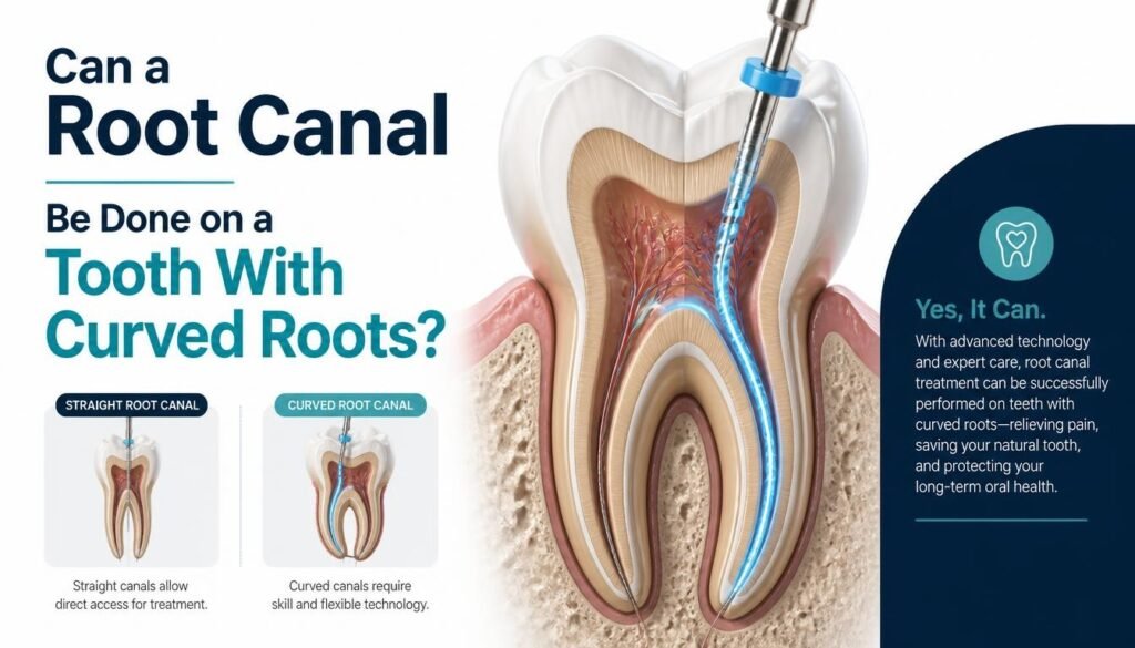
Some root canal cases involve unusual anatomy, curved canals, separated instruments, or other complications.
A dental operating microscope helps provide a clearer view of these challenging areas, supporting the dentist during complex endodontic procedures.
Root Canal Treatment With a Microscope vs. Traditional Treatment
Both conventional and microscope-assisted root canal treatments aim to eliminate infection and preserve the natural tooth.
The difference is primarily in the level of visualization available to the dentist.
| Feature | Conventional RCT | Microscope-Assisted RCT |
|---|---|---|
| Visualization | Direct vision | Magnified vision |
| Illumination | Standard dental lighting | Focused illumination |
| Hidden canals | May be more difficult to identify | Easier to examine |
| Fine anatomical details | Limited visibility | Enhanced visibility |
| Complex cases | Depends on case and clinician | Additional visual assistance |
| Treatment approach | Conventional instruments and techniques | Magnification-assisted precision |
It is important to understand that a microscope does not automatically guarantee a successful root canal treatment. The dentist’s training, diagnosis, experience, infection control, treatment technique, and appropriate restoration all play important roles.
Is Microscope-Assisted Root Canal Treatment Better?
Microscope-assisted root canal treatment can offer valuable advantages, particularly when a tooth has complicated anatomy or requires a high level of precision.
The American Association of Endodontists (AAE) recognizes the usefulness of operating microscopes in endodontics for identifying hidden canals, removing separated instruments, examining complex anatomy, and supporting conservative treatment.
However, the benefits depend on the individual clinical situation. A straightforward root canal may not require the same level of magnification as a complicated retreatment case.
Your dentist will determine whether microscope-assisted treatment is appropriate after examining the affected tooth.
Who May Benefit From Microscope-Assisted Root Canal Treatment?
A dental operating microscope may be particularly useful for patients with:
- Complex or unusual root canal anatomy
- Narrow or calcified root canals
- Previously treated teeth requiring retreatment
- Suspected missed canals
- Persistent infection following earlier treatment
- Teeth with certain cracks or structural defects
- Complicated molar root canal cases
If you have recurring tooth pain or discomfort after a previous root canal, consult a dentist for a detailed evaluation.
Read our guide on how a dentist diagnoses the need for root canal treatment.
Does a Root Canal Microscope Make Treatment Painless?
A microscope is primarily designed to improve visibility and precision. It is not a pain-relieving device by itself.
Root canal treatment is generally performed under local anaesthesia to help keep the procedure comfortable. The level of discomfort during and after treatment depends on several factors, including the condition of the tooth, the extent of infection, and individual pain sensitivity.
Modern dental techniques and appropriate anaesthesia can help make treatment more comfortable for patients.
At The Dental Crowns, our approach to painless root canal treatment in Noida focuses on patient comfort, careful diagnosis, and appropriate treatment planning.
Are There Any Limitations of a Root Canal Microscope?
Although dental microscopes offer several advantages, they also have certain limitations.
Learning and Training
Using a microscope effectively requires specialized training, experience, and practice. The dentist must learn to work comfortably while observing a magnified image.
Treatment Time
Some microscope-assisted procedures may take longer, particularly when treating complex anatomical structures.
Equipment and Cost
Dental operating microscopes require specialized equipment and maintenance. Their availability may vary between dental clinics, and the cost of treatment may depend on the complexity of the case and the technology used.
Not Necessary for Every Case
Every root canal treatment is different. A microscope may provide significant advantages in complex cases, but its use is not essential for every routine procedure.
The most appropriate treatment approach should always be determined through professional dental evaluation.
Why Choose The Dental Crowns for Root Canal Treatment in Noida?
Choosing the right dental clinic is an important step when dealing with tooth pain, infection, or complicated dental problems.
At The Dental Crowns, patients can explore a range of general, restorative, cosmetic, and surgical dental services with a focus on personalized care.
Our approach emphasizes:
- Thorough dental examination and diagnosis
- Patient education and treatment planning
- Modern dental technology
- Focus on preserving natural teeth whenever possible
- Appropriate treatment recommendations based on individual needs
- Comfortable and hygienic dental care
Whether you need a routine root canal or an evaluation for a more complicated dental concern, consulting an experienced dental professional can help you understand your treatment options.
Explore our complete dental services in Noida to learn more.
Frequently Asked Questions (FAQs)
1. What is a root canal microscope used for?
A root canal microscope is used to magnify and illuminate the inside of a tooth during endodontic treatment. It helps dentists examine root canals, identify hidden canals, and perform delicate procedures with improved visibility.
2. How much magnification does a dental microscope provide?
Depending on the model and settings, dental operating microscopes can provide magnification ranging from approximately 4x to 25x, allowing dentists to examine small anatomical structures in greater detail.
3. Can a microscope detect hidden root canals?
A microscope can help dentists identify additional or hidden root canals, particularly in teeth with complex anatomy. However, detection depends on the tooth’s structure and the clinical situation.
4. Is microscope-assisted root canal treatment safe?
Microscope-assisted treatment is an established technique in modern endodontics. Its safety and effectiveness depend on proper diagnosis, professional training, appropriate instruments, and the dentist’s clinical judgment.
5. Does every root canal require a microscope?
No. A microscope may be particularly helpful for complex root canal cases, but many routine root canal procedures can be performed without one. Your dentist will recommend the appropriate approach based on your tooth’s condition.
6. Can a dental microscope help during root canal retreatment?
Yes. Magnification may help identify missed canals, inspect the previously treated area, and assist in removing certain materials or managing complications during retreatment.
7. Is microscope-assisted root canal treatment more expensive?
The cost may vary depending on the clinic, equipment, treatment complexity, and the dentist’s expertise. Ask your dental professional for a personalized treatment estimate before proceeding.
8. Where can I get root canal treatment in Noida?
If you are experiencing tooth pain, sensitivity, or signs of infection, you can consult The Dental Crowns in Sector 79, Noida, for an examination and appropriate treatment recommendations.
Conclusion
A root canal microscope is an important advancement in modern endodontics that helps dentists examine the internal structures of teeth with enhanced magnification and illumination.
From locating hidden canals to assisting in complex retreatment procedures, microscope-assisted dentistry can improve visualization and support precision during treatment.
However, successful root canal treatment depends on several factors, including accurate diagnosis, the dentist’s expertise, appropriate treatment techniques, and proper restoration of the tooth.
If you are experiencing persistent tooth pain or have been advised to undergo root canal treatment, consult a dental professional to understand the most suitable treatment for your condition.
Book your dental consultation at The Dental Crowns, Noida, and take an informed step toward protecting your natural teeth and oral health.