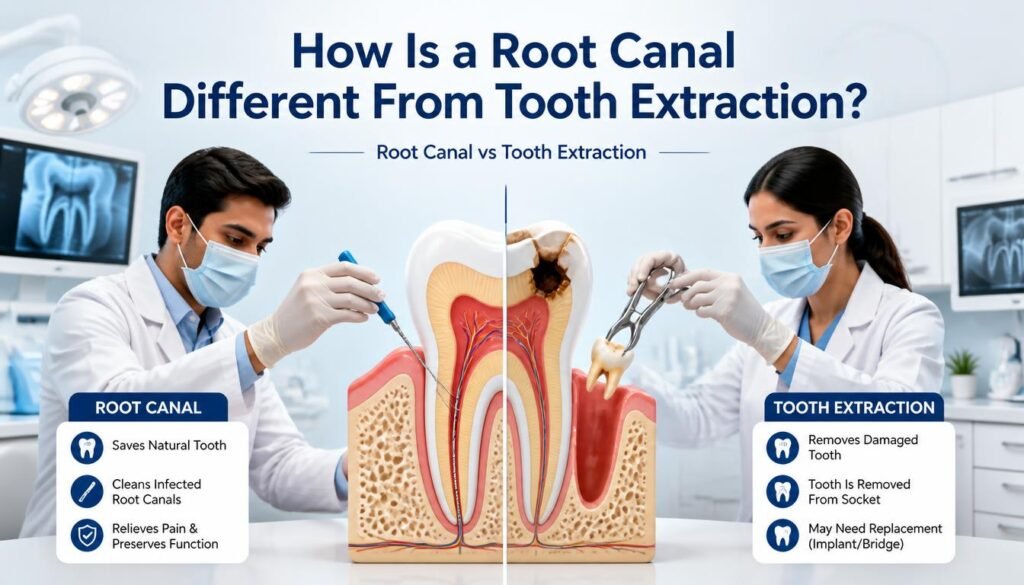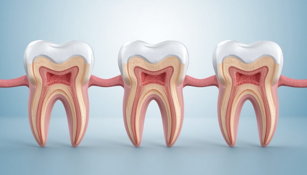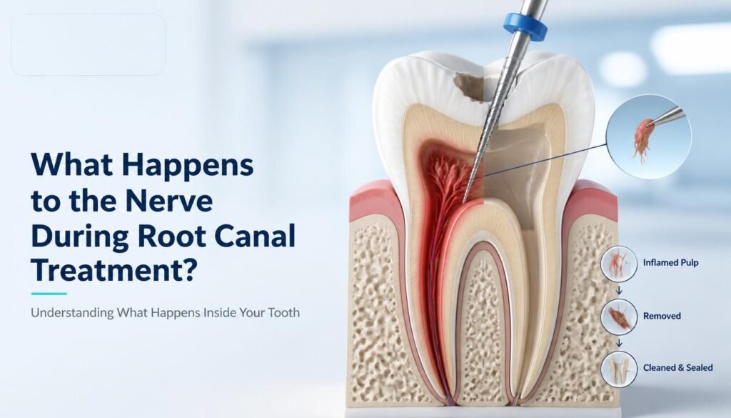

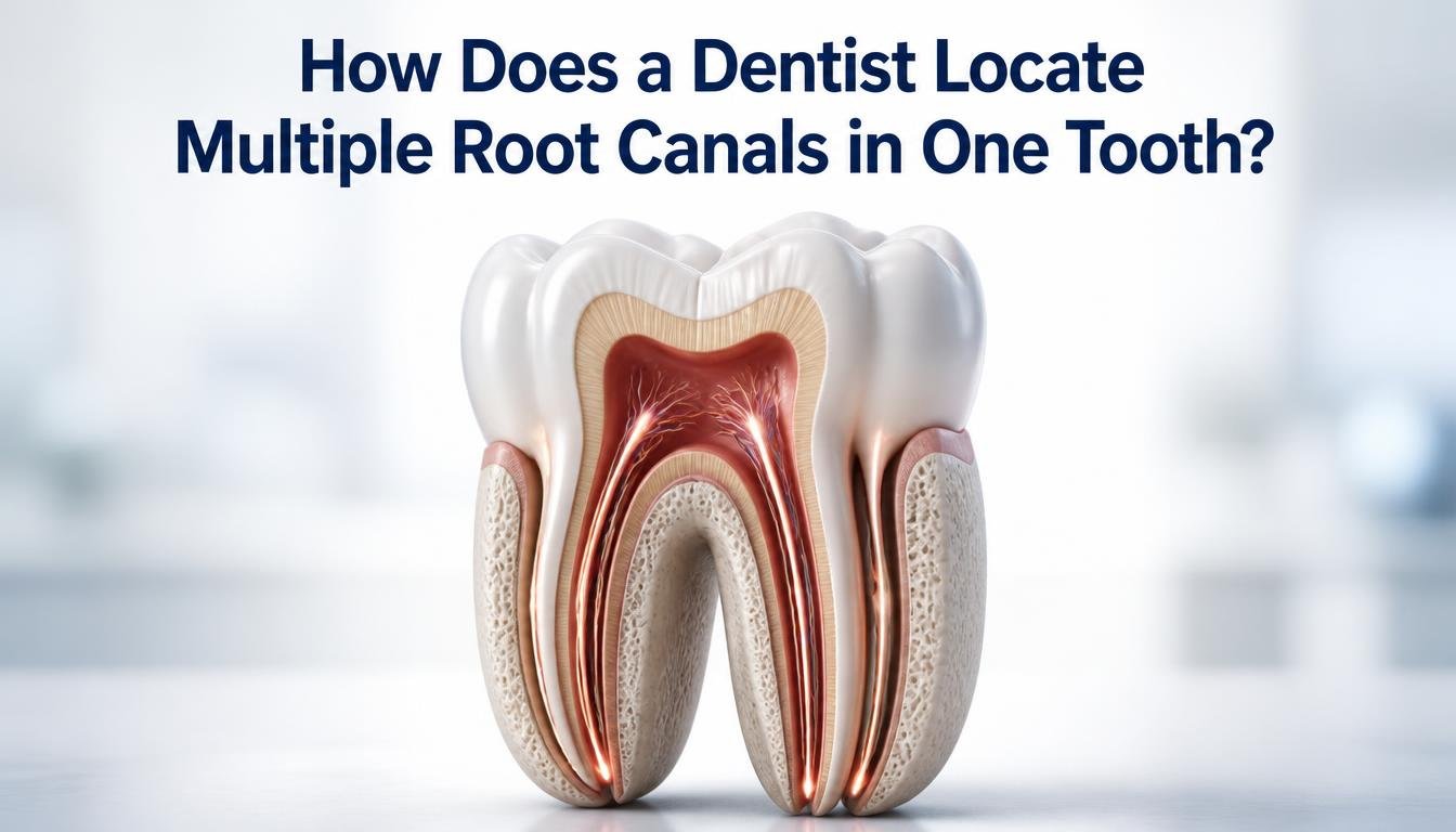
A tooth may look simple from the outside, but its internal root canal system can be surprisingly complex. Some teeth have one canal, while others can contain multiple canals, narrow passages, curved canals, connecting spaces, or additional canals that are difficult to identify during a routine examination.
So, how does a dentist locate multiple root canals in one tooth?
Dentists use a combination of clinical examination, dental X-rays, knowledge of tooth anatomy, careful inspection of the pulp chamber, magnification and, when clinically justified, advanced 3D imaging such as CBCT (Cone Beam Computed Tomography). The goal is to identify the actual canal anatomy so that the dentist can properly clean, disinfect and seal the root canal system.
This is particularly important during complex root canal treatment, because an untreated canal may leave infected or inflamed tissue inside the tooth.
If you are experiencing persistent tooth pain or sensitivity, you can learn more about Painless Root Canal Treatment at The Dental Crowns.
What Does “Multiple Root Canals in One Tooth” Mean?
When people hear the term “root canal,” they sometimes imagine one single tunnel running through the tooth. In reality, the internal anatomy can be much more complicated.
A tooth can have:
- Multiple main root canals
- Additional or accessory canals
- Narrow or flattened canals
- Curved canals
- Connecting spaces between canals
- Lateral canals
- Apical branches
- C-shaped canal configurations
- Partially calcified or narrowed canals
The number and arrangement of canals can vary from tooth to tooth. Even two teeth that look similar externally can have different internal anatomy.
The Dental Crowns’ existing educational content also explains that additional canals and unusual canal configurations can make root canal treatment more technically demanding.
Why Is It Important to Find Every Root Canal?
The purpose of root canal treatment is not simply to clean the largest or easiest canal.
The dentist needs to identify and treat the relevant canal system as completely as possible. If an additional canal containing infected or inflamed tissue is not identified, symptoms can potentially persist or return.
According to the American Association of Endodontists, complicated canal anatomy can be one reason a previously treated tooth may require additional evaluation or retreatment.
This does not mean that every persistent symptom after root canal treatment is caused by a missed canal. Other possibilities include reinfection, leakage, fractures or other dental conditions.
That is why diagnosis is important before deciding on treatment.
How Does a Dentist Locate Multiple Root Canals?
Dentists generally use several methods together rather than depending on a single test.
1. Clinical Examination Comes First
The process usually begins with a detailed dental examination.
The dentist considers:
- Your symptoms
- Location of pain
- Sensitivity to hot or cold
- Pain while biting
- Swelling or tenderness
- Previous dental treatment
- Existing fillings or crowns
- The appearance and condition of the tooth
Clinical tests can help the dentist determine whether the pulp may be inflamed or infected and whether root canal treatment should be considered.
Importantly, symptoms alone cannot tell a dentist exactly how many canals a tooth contains.
2. Dental X-Rays Help Reveal Root Anatomy
A periapical dental X-ray is commonly used to examine a tooth and the surrounding bone.
An X-ray can provide useful information about:
- Number and shape of roots
- Approximate canal direction
- Root curvature
- Bone changes around the root
- Previous root canal treatment
- Other abnormalities
However, conventional dental X-rays are two-dimensional representations of three-dimensional anatomy.
This means that two canals can sometimes overlap on an X-ray, making the internal anatomy more difficult to interpret.
For this reason, dentists may use different angulations or additional diagnostic methods when the anatomy is uncertain.
3. The Dentist Carefully Examines the Pulp Chamber
Once access is made to the inside of the tooth during root canal treatment, the dentist can carefully examine the pulp chamber floor.
The location and shape of the chamber can provide anatomical clues about where canal openings may be located.
A dentist trained in endodontic treatment understands that canal openings are not necessarily positioned randomly. Knowledge of normal anatomy and common anatomical variations helps guide the search.
This is particularly useful when an additional canal is smaller, partially hidden or located in an unexpected position.
4. Magnification Can Help Identify Small Canal Openings
Some canals are extremely small and difficult to see with the naked eye.
Dental magnification can improve visualization of the treatment area and help the dentist inspect the chamber floor and possible canal openings more closely.
In complex cases, magnification may be particularly useful when:
- A canal opening is very small
- The tooth has unusual anatomy
- Calcification has narrowed the canal
- Previous treatment has altered the chamber
- An additional canal is suspected
The purpose is to improve visibility while allowing the dentist to preserve as much healthy tooth structure as possible.
5. CBCT Can Provide a 3D View When Clinically Justified
One of the most useful technologies for difficult root canal anatomy is Cone Beam Computed Tomography (CBCT).
Unlike a conventional dental X-ray, CBCT produces three-dimensional images of the teeth and surrounding structures.
The American Association of Endodontists explains that CBCT can provide valuable additional information in selected endodontic cases, particularly when conventional imaging does not adequately explain the anatomy or pathology.
CBCT may help a dentist evaluate:
- Additional canals
- Complex root anatomy
- Root curvature
- Unusual canal configurations
- Previously untreated anatomy
- Periapical changes
- Certain treatment complications
However, CBCT is not automatically required for every root canal treatment.
The AAE and AAOMR’s updated guidance emphasizes appropriate, case-specific use of CBCT rather than routine scanning without clinical justification.
Therefore, the dentist decides whether the additional information from 3D imaging is useful for the particular patient and tooth.
6. Different X-Ray Angles Can Help Separate Overlapping Canals
Because a conventional dental X-ray is two-dimensional, overlapping structures can sometimes make canals difficult to distinguish.
Taking images from different horizontal angles can provide additional information about the relationship between roots and canals.
This can help the dentist form a better understanding of the three-dimensional anatomy before or during treatment.
The dentist may combine these images with clinical findings and other diagnostic information rather than relying on the X-ray alone.
7. Dental Anatomy Knowledge Plays a Major Role
Technology is helpful, but it is not the only factor.
A dentist’s knowledge of normal and unusual root canal anatomy is important when searching for additional canals.
Different teeth have different typical anatomical patterns, and some teeth are more likely to present with complex configurations.
For example, molars can have multiple roots and canals, while some teeth may have unusual configurations such as C-shaped canal systems.
Knowing where additional canals may commonly occur can help the dentist investigate suspicious areas rather than assuming that the first canal found is the only canal present.
Can a Tooth Have More Root Canals Than Expected?
Yes.
A tooth may have additional canals that are not obvious during an initial examination.
For example, a dentist may expect a particular tooth to contain a certain canal configuration, but further clinical examination or imaging may reveal an additional canal.
The canal may be:
- Very narrow
- Curved
- Partially calcified
- Hidden beneath dentin
- Connected to another canal
- Difficult to distinguish on a conventional X-ray
This is one reason complex root canal treatment requires careful treatment planning.
Why Are Some Root Canals Difficult to Find?
Several factors can make canals harder to locate.
Calcification
Age, previous dental treatment, trauma and other factors can cause mineral deposits that narrow the pulp chamber or canals.
Curved Canals
A canal may curve significantly rather than following a straight path.
Additional Canals
Some teeth naturally contain more canals than expected.
Previous Root Canal Treatment
A previously treated tooth may have altered anatomy, restorations or other changes that make evaluation more complicated.
Crowns and Large Restorations
Existing dental restorations can sometimes make access and visualization more challenging.
Unusual Anatomy
C-shaped canals, connecting spaces, accessory canals and other anatomical variations can require additional planning.
The American Association of Endodontists notes that complex canal anatomy can be a factor when a previously treated tooth does not heal as expected.
What Happens If an Extra Root Canal Is Missed?
A missed canal can potentially leave infected or inflamed tissue inside the tooth.
If symptoms continue after root canal treatment, however, it is important not to assume automatically that a missed canal is responsible.
A dentist or endodontic specialist may investigate several possible causes, including:
- Untreated canal anatomy
- Persistent infection
- New infection
- Inadequate sealing
- Leakage around a restoration
- Tooth fracture
- Other dental or periodontal problems
Additional imaging and clinical examination may be used to determine the actual cause.
Can CBCT Always Find Every Root Canal?
No.
CBCT is a valuable diagnostic tool, but it does not guarantee that every microscopic anatomical feature will be visible.
Image quality can be affected by factors such as:
- Resolution
- Field of view
- Patient movement
- Metal restorations
- Root filling materials
- Imaging artifacts
- The size and complexity of the canal
The American Association of Endodontists describes CBCT as an important adjunct in selected endodontic cases, but imaging still needs to be interpreted alongside the clinical examination.
Does Finding Multiple Canals Make Root Canal Treatment More Difficult?
It can.
The difficulty of root canal treatment depends on factors such as:
- Number of canals
- Canal diameter
- Canal curvature
- Calcification
- Canal connections
- Root anatomy
- Infection
- Previous treatment
- Remaining tooth structure
A tooth with multiple canals does not automatically mean that treatment will be unsuccessful.
The important consideration is whether the dentist can accurately diagnose, access, clean, disinfect and seal the relevant canal system.
Can Laser Dentistry Be Used During Complex Root Canal Treatment?
Laser technology may be used as an adjunct in selected dental procedures, depending on the treatment plan and clinical situation.
It should not be assumed that laser treatment automatically replaces conventional root canal instrumentation.
At The Dental Crowns, laser dentistry is offered as part of its dental services. You can learn more about Advanced Clinical Laser Dentistry at The Dental Crowns.
The appropriate approach depends on the individual tooth, diagnosis and treatment objectives.
When Should You See a Dentist for Possible Root Canal Treatment?
Consider scheduling a dental examination if you experience:
- Persistent toothache
- Severe tooth pain
- Sensitivity to hot or cold
- Pain while chewing
- Swelling around a tooth
- Tenderness
- A recurring dental abscess
- Deep tooth decay
- A cracked or damaged tooth
- Symptoms returning after previous root canal treatment
These symptoms do not automatically mean you need root canal treatment. A professional examination is needed to determine the underlying cause.
How The Dental Crowns Can Help With Root Canal Treatment
The Dental Crowns provides dental services in Sector 79, Noida, including root canal treatment and laser dentistry.
The clinic’s root canal service describes an approach focused on diagnosis, precision and patient comfort.
You can explore:
Frequently Asked Questions
How does a dentist know how many root canals a tooth has?
A dentist uses clinical examination, dental X-rays, knowledge of tooth anatomy and, in selected complex cases, additional imaging such as CBCT to understand the number and configuration of canals.
Can one tooth have multiple root canals?
Yes. Many teeth have multiple canals, and some teeth can contain additional canals beyond the commonly expected anatomy.
Can an extra root canal be hidden?
Yes. An additional canal can be very narrow, curved, calcified or positioned in a way that makes it difficult to identify using a conventional examination or X-ray.
Does every root canal require a CBCT scan?
No. CBCT is not routinely required for every root canal. It may be considered when the additional three-dimensional information is clinically useful for diagnosis or treatment planning.
Can a dentist find a missed root canal after treatment?
A dentist can investigate a previously treated tooth when symptoms persist or return. Clinical examination, conventional imaging and, when appropriate, CBCT may help identify untreated anatomy or other possible causes.
Are molars more likely to have multiple root canals?
Molars can have multiple roots and canals and may present with complex anatomy. The exact number and configuration vary between individual teeth.
Is a root canal with multiple canals more painful?
The number of canals does not by itself determine how much pain a patient experiences. Modern root canal treatment is generally performed with local anaesthesia, while post-treatment soreness can vary between patients and procedures.
What happens if a dentist cannot locate a canal?
If a canal is difficult to locate, the dentist may use additional magnification, imaging, anatomical knowledge and specialized techniques. For particularly complex cases, referral to an endodontic specialist may also be considered.
Final Takeaway
How does a dentist locate multiple root canals in one tooth?
The answer involves a combination of diagnostic methods rather than one single technique. Dentists may use clinical examination, dental X-rays, different imaging angles, careful inspection of the pulp chamber, magnification and, when clinically justified, CBCT 3D imaging.
The objective is to understand the individual tooth’s anatomy as accurately as possible so the relevant root canal system can be properly treated.
If you have persistent tooth pain, sensitivity, swelling or symptoms after previous root canal treatment, a professional dental examination can help determine whether complex canal anatomy or another dental problem may be involved.